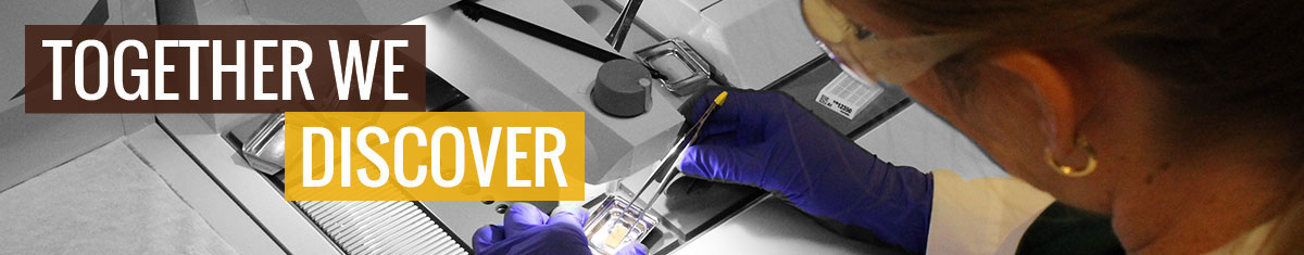
The Research Histology Laboratory provides an extensive array of routine and specialized histological services, as well as immunohistochemical stains. The laboratory boasts highly skilled and quality technical and professional pathology services and is capable of assisting researchers at WMed and beyond. Take a look at the laboratory’s full list of services:
Routine Histology
Tissue trimming, processing, embedding, sectioning and staining for routine and research samples.
- Paraffin and frozen sectioning and staining
- Bone decalcification and sectioning
- Unstained slides, serial and step sectioning
- Conventional staining
- Barcode labeling of cassettes and slides
Specialty Histology Staining
Histochemical techniques to demonstrate various tissue components.
|
|

Immunohistochemistry
Immunohistochemistry for both routine and novel markers available at WMed include, but are not limited to:
|
|
Specialty Services
- Sledge Microtome
- 2" x 3" Macrosette paraffin processing of decalcified large bone specimen or soft tissue.
- Frozen sectioning of large soft tissue up to 4”x5”.
- Free-floating Immunohistochemistry
Digital Pathology
- Slide scanning 20x, 40x with the Leica Aperio system.
- Image analysis provides quantifiable data for a variety of parameters.
Additional stains, antibodies, or custom requests are welcome. For more information, please contact Kristi Bailey, BS, HTL, Lead Histotechnologist at kristi.bailey@wmed.edu.