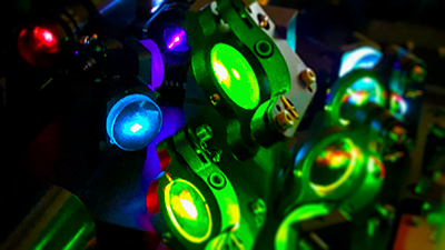
The Flow Cytometry and Imaging Core provides state-of-the-art equipment and expertise in flow cytometry, cell sorting, and imaging. Core Manager Michael Clemente, MS, SCYM(ASCP), a flow cytometrist with extensive experience in immunology and hematology/oncology research, is available for project/experimental design consultation, data analysis, instrument setup, full-service cell sorting, and training. Comprehensive one-on-one training is available to enable independent operation of flow and imaging instruments by investigators and staff.

Flow Cytometry and Cell Sorting Instruments
Analyzers
Attune Nxt Analyzer, ThermoFisher Scientific
16-parameter (14 color) instrument equipped with 4-lasers (488, 640, 405, 561) and a 96 well plate loader for high throughput experiments.
Fortessa Special Order Research Product (SORP) Analyzer, BD Biosciences
20-parameter (18 color) instrument equipped with 5-lasers (488, 405, 640, 355, 561).
Aurora Analyzer, Cytek Biosciences
5-laser (355, 405, 488, 561, 640) spectral instrument with a versatile Automated Sample Loader for high throughput experiments.
Sorters

Influx Cell Sorter, BD Biosciences
16 parameter (14 color) instrument equipped with 4-lasers (488, 640, 405, 355) and 4-way sorting, housed in a Baker BioProtect IV Class II Type A2 biosafety cabinet
Aurora CS Cell Sorter, Cytek Biosciences
5-laser (488, 640, 405, 355, 561) spectral instrument capable of 6-way sorting, housed in a Baker BioProtect IV Class II Type A2 biosafety cabinet
Melody Cell Sorter, BD Biosciences
User friendly 11 parameter (9 color) instrument equipped with 3-lasers (488, 405, 640) and 4-way sorting
Microscopy/Imaging Instrumentation
A1R+ Confocal Microscope System, Nikon
Laser-scanning confocal microscope equipped with 405, 488, 561, and 640nm lasers. Galvano scanner enables high-resolution imaging, resonant scanner allows for fast imaging, and the high speed Z-stage allows for efficient Z-stack images. Live cell imaging is available with a CO2 and temperature control system. NIS-Elements C control software enables integrated control of the confocal imaging system, microscope, and peripheral devices with a simple and intuitive interface.
Lionheart FX Automated Live Cell Imager, Biotek
Easy to use, versatile LED based automated fluorescent and phase contrast microscope with accessible filter cubes adaptable for detection of a variety of fluorochromes.
EVOS M7000, ThermoFisher Scientific
Epifluorescent microscope capable of 4 color and phase contrast images and a wide variety of vessel types for imaging.
FLoid, ThermoFisher Scientific
Located in our tissue culture core, the FLoid is single objective basic 3 color fluorescent microscope with digital zoom, allowing investigators quick assessment of fluorescent intensity

