The cases come across their desks often, death investigations involving skull fractures in adults and children, some of them with the same nagging question: When did the trauma occur?
For forensic practitioners, the exact answer, unfortunately, has often proven scientifically elusive.
Now, with the help of a two-year, $576,000 grant from the National Institute of Justice (NIJ), two anthropologists and a pathologist from WMed are taking an important first step to try to change that.
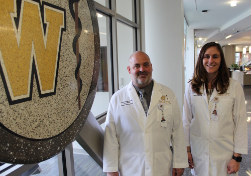
Carolyn V. Isaac, PhD, Jered B. Cornelison, PhD, and Joseph A. Prahlow, MD, each of whom are faculty in the medical school’s Department of Pathology, are co-principal investigators on the project, “Investigations on the Cellular and Morphologic Characteristics of Cranial Vault Fracture: Research and Development of a Time Since Fracture Protocol and Database.” The co-investigators include Rudolph Castellani, MD, Amanda Fisher-Hubbard, MD, Wendy Lackey, PhD, and Brandy Shattuck, MD, all faculty in the Department of Pathology, as well as Joyce deJong, DO, chair of the Department of Pathology,
Dr. Isaac said the impetus for the study stemmed from forensic cases, especially potential child abuse cases, analyzed by her and Drs. Cornelison and Prahlow where there was evidence of skull fractures in different stages of healing.
“The big question was, ‘When did they happen?’” Dr. Isaac said. “And does it corroborate the stories we were getting from the caregivers?”
Dr. Isaac said she and her colleagues began digging, initially assuming that previous research existed on how to determine the timing of when a fracture had occurred. What they found, however, were references that were based only on anecdotal evidence, radiographic studies that did not include samples representing the entire age range from infant to adult, and virtually nothing on how cranial fractures heal.
Given those results and recognizing what they saw as a significant gap in forensic science, Drs. Isaac, Cornelison and Prahlow, in February, sought funding from the NIJ, the research, development and evaluation agency of the U.S. Department of Justice. The grant was awarded on September 29, 2017, and the funds will officially become available on January 1, 2018.
The research by Drs. Isaac, Cornelison and Prahlow will center on the creation of a database of decedents with skull fractures of known injury dates with additional information on the cause of the fractures, co-morbidities and photographic, radiologic and histological documentation of the injuries. Their research will also focus on the histological evaluation of skull fracture healing to determine the cellular and tissue progression at different anatomic zones within the fracture sites and establish stages of cranial fracture healing with specific tissue and cellular characteristics.
To build what they hope will be a robust database of decedents, Drs. Isaac and Cornelison said that, as part of the study, they are partnering with six to seven medical examiner offices from across the U.S. to collect samples from cases in which the mechanisms and dates of previous injuries are known.
The fracture samples will be sectioned and treated with five different histochemical stains as part of the project to optimize the visibility of the cellular and tissue healing response.
“The study is going to look at the microscopic anatomy of those healing fractures and doing a pathological and histological assessment and a description of what is seen,” Dr. Isaac said. “We want to look and see what the pattern is when cells are coming in and exiting again and see what cells are apparent at different time stages to understand how does fracture healing proceed.”
Dr. Cornelison said the project would not be possible without the unique position WMed has in the forensic community. He said the combination of forensic pathologists, forensic anthropologists, and the specialized facilities at the medical school, including histology, biostatistics, and the Office of the Medical Examiner, creates the perfect environment to conduct this research.
Dr. Isaac and Dr. Cornelison are hopeful that the two-year project will lead to a better understanding of how skull fractures heal and become a national model for the sampling, analysis and evaluation of healing skull fractures.
They also are hoping to set the stage for further study and, eventually put in place a way to scientifically determine the age of healing skull fractures.
“Right now, it’s not common practice for folks to take samples of healed fractures and look at them histologically,” Dr. Isaac said. “Our hope is that people will start putting this into practice and eventually we will have enough data to empirically answer the question, ‘When did this fracture occur?”
