
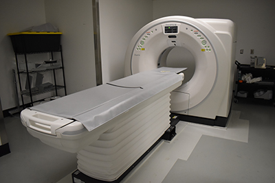
A new piece of equipment at WMU Homer Stryker M.D. School of Medicine (WMed) is greatly improving the efficiency -- and expanding research opportunities -- within the Department of Pathology.
The new Fujifilm Supria Plus 16-Slice CT Scanner arrived at the W.E. Upjohn M.D. Campus in March, allowing forensic pathologists access to high-quality diagnostic imaging.
It’s an incredibly valuable asset for the department, which serves as the Office of the Medical Examiner for 14 Michigan counties and provides forensic science consultant services for several additional counties in Michigan and northern Indiana.
“This opens a whole new avenue for WMed in terms of clinical application but also research,” said Christine Pink, PhD, a forensic anthropologist and assistant professor in the Department of Pathology. “We're one of very few medical examiner offices in the country that has a CT scanner. This allows us to go above and beyond best practice, and we'll have the opportunity to collect data to demonstrate the efficiency of postmortem CT imaging.”
The Office of the Medical Examiner and Forensic Services receives approximately 1,600 decedents each year, more than half of whom require a full autopsy, according to Dr. Pink. The new CT scanner -- with its ability to provide detailed, cross-sectional images of bones, organs, and soft tissues -- greatly streamlines the postmortem examination process.
“There are a lot of things our pathologists can diagnose via CT imaging,” Dr. Pink said. “We’re able to do these scans quickly, alleviating a huge burden on our technicians. Given the volume of cases we see each year, having another tool in our repertoire to be able to streamline things is extremely valuable.”
The new scanner produces research quality imaging data, expanding the research possibilities at WMed at a time when postmortem imaging – and CT scans in particular – continues to grow as an area of interest.
Shamsi Berry, PhD, MS, associate professor and vice chair of the Department of Biomedical Informatics, is currently working with the Department of Pathology to secure extramural grant funding to support research using postmortem CT imaging data.
“We're very excited to make people aware that we have this tremendous resource,” Dr. Pink said. “If you can dream it, we can probably figure out how to do it research-wise. Once we have some extramural funding, we’ll have the capability on a larger scale to make this imaging data available for research.”